Latest Version
Version
1.2.1
1.2.1
Update
December 04, 2024
December 04, 2024
Developer
PlumpTurtle Creative Studio
PlumpTurtle Creative Studio
Categories
Education
Education
Platforms
Android
Android
Downloads
0
0
License
Free
Free
Package Name
com.plumpturtlecreativestudio.parasitology_flashcard
com.plumpturtlecreativestudio.parasitology_flashcard
Report
Report a Problem
Report a Problem
More About Human Parasitology Flashcard
Microscopy identification is still regarded as the gold standard for laboratory diagnosis of human parasitic diseases.
Due to the above reason, this app is created to facilitate the learners to be familiarised with the key microscopic characteristics of human parasites as observed in various specimens, including faecal material, blood, body fluid or tissue.
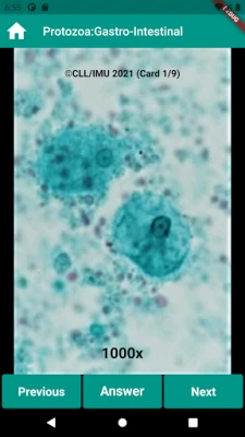
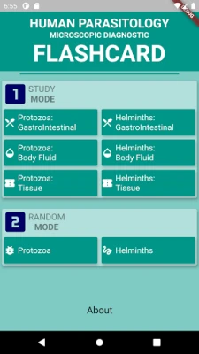
The author has included a total of 80 microscopic images of the common human parasites. These parasites are divided into 2 clusters, namely protozoa and helminth; each is further sub-divided into 3 groups which are the gastrointestinal, blood & body fluid and tissues specimen.
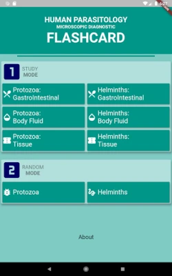
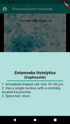
Two different modes can be utilised by the learners, depending on their learning needs, either the study mode or the random mode. In the study mode, the learners can learn by going through the properly-structured flow of images and answers, in which the answers have included the scientific and common name of the parasite, and the description of the key morphology characteristic. The learners can also attempt the random mode, in which an image will be randomly shown to knowledge check purpose.
Rate the App
Add Comment & Review
User Reviews
Based on 0 reviews
No reviews added yet.
Comments will not be approved to be posted if they are SPAM, abusive, off-topic, use profanity, contain a personal attack, or promote hate of any kind.
More »


Popular Apps
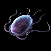
Human Parasitology FlashcardPlumpTurtle Creative Studio

FlipaClip: Create 2D AnimationVisual Blasters LLC

Medical Parasitology Lab.CoderBot Ltd.
Parasitology Picto QuizDr. Arunava Kali

KingDraw: Chemistry StationPrecision Agriculture technology Co.,L.td

Wombat: Play, Earn, ConnectSpielworks

Time Planner: Schedule & TasksOleksandr Albul

Devikins: RPG/ NFT/Crypto GameMoonLabs Game Studios LTDA

Matty the Water Molecule GameEngaging Every Student

Molecular geometry - MirageM. Chardine
More »


Editor's Choice

Panda Video Compress & ConvertFarluner Apps & Games

Like A Dino!super_toki

To-Do List - Schedule PlannerDairy App & Notes & Audio Editor & Voice Recorder

Game of Empires:Warring RealmsDroupnir Entertainment Limited.

Startup Empire - Idle TycoonLittleBit!

Parental Control App- FamiSafeShenzhen Wondershare Software Co., Ltd.

Monitor & ControlSony Corporation

MMGuardian Parental ControlMMGuardian.com

Google DocsGoogle LLC

Document Reader - PDF EditorSimple Design Ltd.
